Is My Ankle Broken or Sprained? How to Tell
Foot Care Guide from ASG Foot & Ankle
The short answer
How to tell if an ankle is broken or sprained: only imaging can confirm it. Clinicians order an X-ray if the bone is tender at four set spots or you cannot take four steps. When none apply, a large review puts fracture risk under 1.4% (Bachmann 2003). When one applies, only about 1 in 5 ankles is broken.
- Bone through the skin, ankle out of line, or a cold, pale or numb footThis needs emergency care whatever the injury turns out to be. Go to the ER now.
- Bone tender at one of the four spots, or you cannot take four stepsThe rule says X-ray. Roughly 4 in 5 of these ankles are not broken. Get an X-ray today.
- Diabetes, neuropathy or reduced feeling in the footLess feeling can hide a fracture, so the rule cannot clear you. Get it examined.
- None of these, and the injury is under 2 days oldA fracture is unlikely, though not impossible. Protect it and re-check in a few days.
ASG Foot & Ankle patient education. Updated November 6, 2026. General information, not a diagnosis.
Is my ankle broken or sprained? A quiz built on the Ottawa Ankle Rules
Press each spot on the bone with one finger and answer for today. Spots 1 to 4 are shown in the pictures just below the questions. If you see bone, or the foot is cold, pale or numb, skip this and get emergency care.
- 1. Is the back edge or tip of your outer ankle bone tender when you press it?Spot 1. Press on the bone along the back edge of its lowest 6 cm (about 2.4 inches), then on its tip. Soft tissue around the bone does not count.
- 2. Is the back edge or tip of your inner ankle bone tender when you press it?Spot 2. Same check on the inner side: the back edge of the lowest 6 cm, then the tip.
- 3. Is the base of your fifth metatarsal tender when you press it?Spot 3. The bony bump about halfway along the outer edge of your foot.
- 4. Is your navicular bone tender when you press it?Spot 4. The bony bump on the inner side of your foot, in the arch, in front of the ankle.
- 5. Were you unable to take four steps right after the injury, and are you still unable to now?Answer Yes only if both are true. A limp still counts as a step if you complete all four. Do not force it.
- 6. Is a bone showing through the skin?A bad cut or open wound over the injury also needs emergency care.
- 7. Does your ankle look obviously out of line or sit at an odd angle?
- 8. Is your foot cold or pale, or are your toes numb, blue or white?
- 9. Do you have diabetes, neuropathy or reduced feeling in your feet?Reduced feeling can hide tenderness, so the rule cannot clear you.
- 10. Does any of these apply: you are pregnant, the injured person is a child, you have been drinking or using drugs, you hit your head, or you have other painful injuries?The rule's studies or the guidelines that use it leave these situations out.
- 11. Did this injury happen more than 2 days ago?Most studies checked people within 48 hours. A Yes does not change the result below, but read a reassuring result with more caution.
Answer each question. 0 of 11 done.
This check does not diagnose anything. The Ottawa Ankle Rules were written for trained clinicians examining adults, and they were not designed as a self-test. In one study, patients and clinicians agreed poorly when scoring the same ankle. If you are worried, get seen whatever the result says.
X-ray already clear? Ankle sprain recovery timeline
X-ray shows a break? Ankle fracture recovery timeline
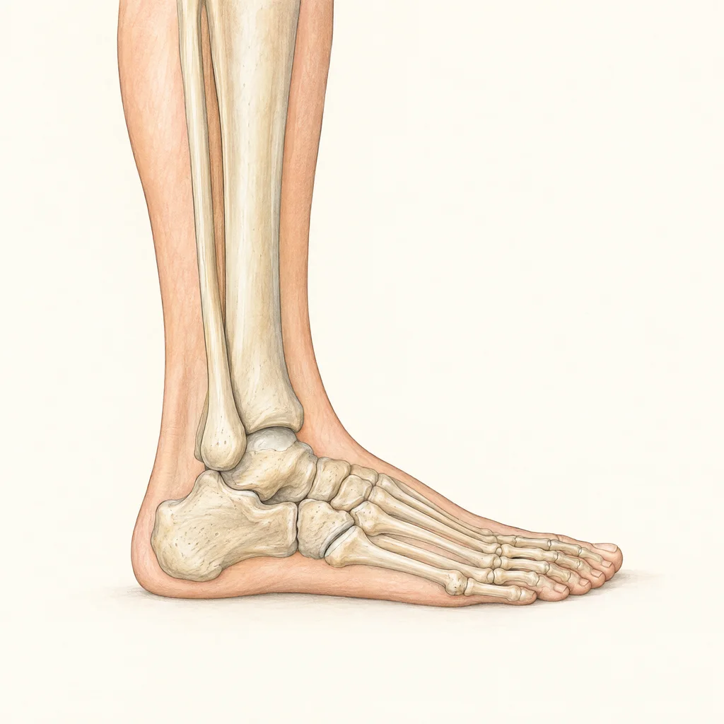
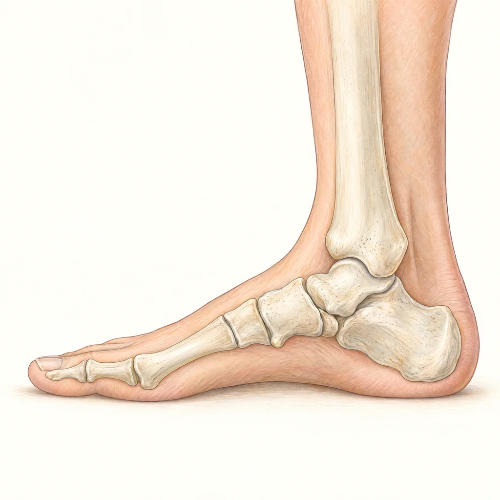
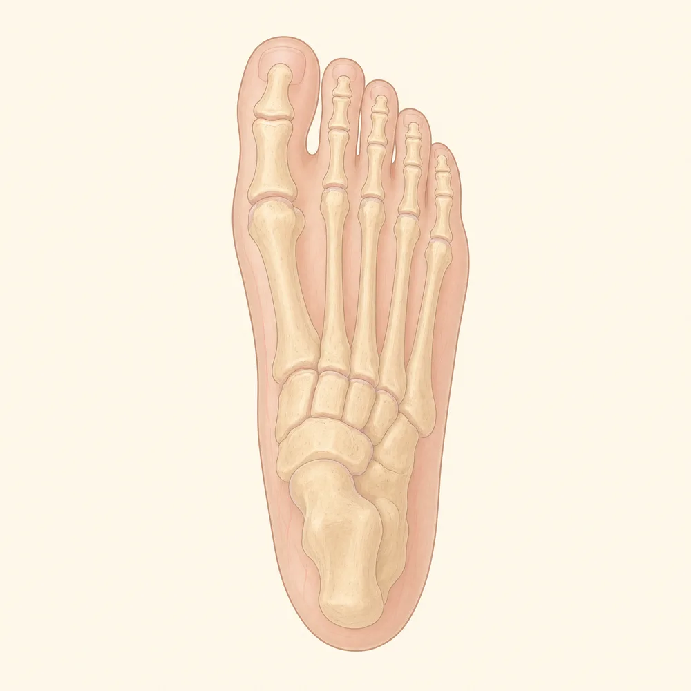
Where to press: the four spots in the Ottawa rule
The rule asks about bone tenderness at four exact places. In a typical sprain, the soft tissue over the outer ligaments is tender (AAFP 2012), so that pain does not count here.
Spots 1 and 2, with pain around the ankle bones, call for an ankle X-ray. Spots 3 and 4, with pain in the midfoot, call for a foot X-ray. Being unable to take four steps counts for either area (NHS Scotland).
 Illustration
- 1Outer ankle bone. The lateral malleolus, the lower end of the fibula. Check the back edge of the lowest 6 cm (about 2.4 inches), then the tip.
- 3Base of the fifth metatarsal. The bony bump about halfway along the outer edge of the foot, where the bone to the little toe begins.
 Illustration
- 2Inner ankle bone. The medial malleolus, the lower end of the tibia. Check the back edge of the lowest 6 cm (about 2.4 inches), then the tip.
- 4Navicular. The bony bump on the inner side of the midfoot, in the arch, in front of the ankle.
 Illustration
- 3Base of the fifth metatarsal. On the outer edge of the foot.
- 4Navicular. On the inner side of the foot.
How accurate are the Ottawa Ankle Rules?
Sensitivity is the share of real fractures the rule flags. Specificity is the share of unbroken ankles it clears. Numbers in brackets are 95% confidence ranges. These figures all come from clinicians using the rule.
| Study | Sensitivity | Specificity |
|---|---|---|
| Validation study: 453 adults, 69 fractures (Stiell 1993) | 100% (ankle 93 to 100, foot 83 to 100) | Not in abstract |
| Review: 27 studies, 15,581 patients (Bachmann 2003) (AAFP 2003) | 97.6% (96.4 to 98.9) | 31.5% (23.8 to 44.4) |
| Review: 66 studies, ankle rule (Beckenkamp 2017) | 99.4% (97.9 to 99.8) | 35.3% (28.8 to 42.3) |
| Review, adults only: 15 studies, 8,560 adults (Gomes 2022) | 91% (89 to 92) | 25% (24 to 26) |
| Review, children: 12 studies, 3,130 children (Dowling 2009) | 98.5% (97.3 to 99.2) | Not in abstract |
The 2022 adult review came in lower than the others. Its authors say three studies pulled the figure down: a military-only group, a study with changed criteria and a retrospective audit (Gomes 2022). The 2017 review also found lower accuracy in its low-bias studies and in studies that X-rayed everyone (Beckenkamp 2017). A negative result lowers the risk a lot without removing it.
The first study did not count avulsion flakes of 3 mm or less as significant fractures (CanadiEM on Stiell 1992), so a tiny chip can sit behind a negative result.
How many X-rays the rule avoids
- Predicted: 34% fewer ankle and 30% fewer foot X-ray series (Stiell 1993), or 30 to 40% fewer unnecessary X-rays overall (Bachmann 2003).
- Measured in a two-hospital trial (2,342 adults): 28% fewer ankle and 14% fewer foot X-rays where the rule was used, while the comparison hospital rose 2% and 13%. No fractures were missed (Stiell 1994).
- Measured at 8 hospitals (12,777 adults): ankle X-ray use fell from 82.8% to 60.9%. Fractures found after discharge stayed about the same, at 0.5% before and 0.4% after (Stiell 1995).
- Children: 24.8% fewer X-rays, ranging from 5% to 44% across studies (Dowling 2009).
Good at ruling a fracture out, poor at ruling one in
A positive result means you should get an X-ray. It does not mean the ankle is broken. Bone tenderness and trouble walking are common with sprains too, so most ankles the rule flags turn out fine. Under 15% of people X-rayed for an ankle sprain have a fracture (Bachmann 2003).
Worked example: 100 injured ankles, 15 of them broken
About 15 have a fracture and about 58 do not. That is roughly 1 in 5.
Fewer than 1 has a fracture. The review's own figure is under 1.4%.
Red squares are fractures. This page did the arithmetic from 97.6% sensitivity, 31.5% specificity and the 15% fracture rate the review assumed (AAFP 2003). The lower 2022 adult figures (Gomes 2022) give about 6 fractures per 100 negative results and about 1 in 6 among positive results.
Who the Ottawa rule was not tested on
The original adult studies left out people under 18, pregnant patients, skin-only injuries, injuries over 10 days old, return visits and people referred in with X-rays (EBM Consult). Guidelines that use the rule add reduced feeling, intoxication, head injury and multiple painful injuries (NHS Scotland). Self-checks are a further gap: a study of people over 15 found poor agreement between patients and clinicians (Blackham 2008).
Reduced feeling, neuropathy or a nerve disorder
The American College of Radiology says a person with neuropathy does not meet the rule's requirements (ACR 2020). The rule depends on pain, and people with reduced feeling may keep walking on an injury and make it worse (ACFAS). Get examined. A hot, red, swollen foot needs prompt review for Charcot foot.
Alcohol or drugs
Your answers about pain and walking may not be reliable. Have the ankle examined, and re-checked once sober.
Several painful injuries, or a head injury
Worse pain elsewhere can hide ankle tenderness, and a head injury can make the check unreliable. Get examined.
Pregnancy
The rule has no tested result for you. Tell staff if you are or might be pregnant, and the doctor will weigh the risks before offering an X-ray (NHS).
Children, especially young children
Later work in children over 5 found 98.5% sensitivity, with 10 of 671 fractures missed (Dowling 2009), and the rule was less sensitive in children than in adults (Beckenkamp 2017). Guidance differs: the ACR accepts age 5 and up (ACR 2020), NHS Scotland excludes under 18, and data are limited under age 6 (Radiopaedia). It only applies to a child who could already walk independently (Carvalho 2025). A child who cannot say where it hurts needs an exam.
An injury that is days old
The rule is mostly used within 48 hours (Gomes 2022), and sensitivity was slightly lower after that (AAFP 2003). The ACR does apply it to pain lasting up to 3 weeks (ACR 2020). A clean result on day 5 tells you less than one on day 1, so an ankle that still hurts after several days needs an exam.
Sprained ankle vs broken ankle: signs that do not sort them
Without an X-ray, it can be very hard to separate a fracture from a severe sprain unless the ankle is visibly out of shape (Patient.info). Gross deformity should not occur with a sprain (AAFP 2001).
"I can walk on it, so it is not broken"
A simple break in one bone may not stop you from walking (AAOS). In a study of 200 people at 3 emergency departments, 4 of the 34 fractures were in people who could take four steps (Amiri 2017). Most of those injuries came from vehicle crashes, so this is one small data point. Putting weight on some fractures too early can move the pieces (AAOS).
"I cannot walk on it, so it must be broken"
In the same study, 64 of the 94 people who could not take four steps had no fracture (Amiri 2017). A complete ligament tear can also stop you walking (Cleveland Clinic).
"I can wiggle it, so it is not broken"
A person may have a broken bone even if they can move the limb (British Red Cross). When doctors examined the same ankle, they agreed poorly on range of motion, with a kappa of .33 (1 is perfect agreement, 0 is chance) (Stiell 1992). It is not part of the rule. Toes still need a look: numb, blue or white toes need emergency care (NHS).
"I heard a pop or a crack, so it is broken"
A snap does not separate a sprain from a fracture (Patient.info), and a severe ligament tear can cause a pop you hear or feel (AAOS). Hearing no sound does not clear the ankle either, because sound plays no part in the rule (NHS Scotland).
"The bruise is huge or very dark, so it is broken"
Bruising and swelling are common with sprains (AAOS), and marked bruising may mean a complete ligament tear or a fracture (Patient.info). Doctors agreed poorly on bruising (kappa .39) and on swelling over the main outer ligament (.18), compared with .66 to .78 for selected bone spots and .83 for ability to bear weight (Stiell 1992). Color mostly shows age: a bruise moves from pinkish red through blue to greenish yellow as it heals (MedlinePlus). It can also drift into the foot and toes with gravity (AAFP 2012).
What an X-ray can miss, and the re-check at about 6 weeks
X-rays show bone (AAOS), and several foot and ankle fractures are frequently missed at the first exam (AAFP 2002).
- Talar dome injury (cartilage and bone on top of the talus). Symptoms are similar to a sprain and often occur with one, and first films may look normal (AAFP 2002). It may take 2 to 4 weeks to show on X-ray (AAFP 2001). MRI found an acute lesion on the outer talar dome in 20 of 261 sprained ankles (7.7%) in athletes (Roemer 2014). A loose fragment can cause locking and swelling (AAFP 2002).
- Lateral process of the talus (snowboarder's fracture). 33 to 41% are missed at the first visit. The tender point sits in front of and below the outer ankle bone (AAFP 2002).
- Back of the talus, and the front of the heel bone. In one series, 17 of 20 patients with a posterior talus fracture were first told they had a sprain. Fractures of the front of the heel bone are hard to assess on routine X-rays, and the pain rises with walking (AAFP 2002).
- Hairline or stress fracture. The crack may not show for several weeks. MRI or a bone scan can find it earlier (AAOS). Small stable breaks have their own course: minor ankle fracture recovery.
- A break high on the fibula. The upper fibula is on the list of commonly missed fractures (Patient.info), and it can break when you sprain your ankle (Cleveland Clinic). None of the four spots checks it, so mention any pain higher up the leg. Pain just above the ankle may be a high ankle sprain.
Why an ankle not improving at about 6 weeks gets re-checked
Most sprains settle in 2 to 6 weeks (AAFP 2012). If one still causes symptoms after more than 6 weeks, CT or MRI should be considered to look for a talar dome lesion (AAFP 2001). AAOS says pain past 4 to 6 weeks may mean a chronic sprain, with MRI an option past 6 to 8 weeks (AAOS).
No single date is agreed. The American College of Radiology rates MRI or CT as usually appropriate for pain lasting 1 to 3 weeks after a negative X-ray (ACR 2020). The 2025 AAFP review advises an X-ray if none was done, or MRI if one was negative, when recovery is off course (AAFP 2025). An ankle that is slowly improving can reasonably wait for the 6-week mark. One with severe pain and no improvement should be seen sooner.
Once you know which injury it is
- Ligament stretched: Grade 1 ankle sprain recovery
- Ligament partly torn: Grade 2 ankle sprain recovery
- Ligament fully torn: Grade 3 ankle sprain recovery
- Tender at spot 3: Fifth metatarsal fracture recovery
- Pain in the middle of the foot: Foot sprain recovery time
The bottom line
- You usually need an X-ray or other imaging to know for sure whether an ankle is broken.
- The Ottawa rule is very good at clearing an ankle and poor at confirming a break, so a positive result means get an X-ray.
- Walking, wiggling, a pop and the size or color of a bruise do not sort a break from a sprain.
- The rule cannot clear you if you have reduced feeling, are intoxicated, have other painful injuries or are pregnant, and it needs caution in young children and in injuries that are days old.
- A normal X-ray can miss cartilage damage and small fractures, so an ankle not improving by about 6 weeks gets re-checked, sooner if the pain is severe.
Frequently Asked Questions
Can my ankle be broken and still walk on it?
Yes. A simple break in one bone may not stop you from walking, and many isolated fractures of the outer ankle bone are stable enough to bear weight. In one study of 200 people, 4 of 34 fractures were in people who could take four steps. Walking can still move some fractures out of place, so get an X-ray if a bone spot is tender.
Can you wiggle a broken ankle?
Often, yes. A person can have a broken bone and still move the limb, so movement does not clear an ankle. Range of motion was one of the least reliable findings when doctors examined the same ankle, and it is not part of the Ottawa rule. Toes are still worth checking, because numb, blue or white toes need emergency care.
Can you still walk on a sprained ankle?
Often, yes, though it may hurt. A grade 2 sprain may be painful to walk on and feel unstable, and a complete ligament tear may stop you walking at all. Clinicians often encourage weight bearing as tolerated once a fracture is not a concern. If you could not take four steps right after the injury and still cannot, get an X-ray first.
How can you tell if an ankle is broken without an X-ray?
You usually cannot be sure. An ankle that is clearly out of line, or a bone through the skin, needs emergency care. Otherwise the Ottawa rule estimates whether an X-ray is needed by checking four bone spots and whether you can take four steps. A negative result lowers the odds of a fracture, but the rule cannot confirm one.
What is the difference between a hairline fracture and a sprain in the ankle?
A hairline or stress fracture is a small crack in a bone. A sprain is a ligament injury. Both can swell and bruise. Stress fracture pain usually builds with activity, eases with rest and is tender over the bone. The crack may not show on X-ray for several weeks, so an MRI or bone scan may be needed if suspicion remains.
Sources and further reading
- Stiell 1992: Interobserver agreement in the examination of acute ankle injury patients
- CanadiEM summary of Stiell 1992: the study that developed the rule
- Stiell 1993, JAMA: Refinement and prospective validation
- Stiell 1994, JAMA: Implementation of the Ottawa ankle rules
- Stiell 1995, BMJ: Multicentre trial of the Ottawa ankle rules
- Bachmann 2003, BMJ: Systematic review of the Ottawa ankle rules
- AAFP 2003: Summary of the Bachmann review
- Dowling 2009: Meta-analysis in children
- Beckenkamp 2017, BJSM: Systematic review with meta-analysis
- Gomes 2022, BMC Musculoskeletal Disorders: Meta-analysis in adults
- Amiri 2017: The four-step test used alone
- Blackham 2008, Emerg Med J: Can patients apply the Ottawa ankle rules to themselves?
- Carvalho 2025: Ottawa rules in children and adolescents
- Roemer 2014, Am J Sports Med: MRI study of acute ankle sprains in athletes
- NHS Scotland emergency guideline: Ottawa Ankle Rules
- American College of Radiology 2020: Acute Trauma to the Ankle
- AAFP 2001: Management of Ankle Sprains
- AAFP 2002: Foot Fractures Frequently Misdiagnosed as Ankle Sprains
- AAFP 2012: Update on Acute Ankle Sprains
- AAFP 2025: Management of Acute Ankle Sprains
- AAOS OrthoInfo: Ankle Fractures
- AAOS OrthoInfo: Sprained Ankle
- AAOS OrthoInfo: Stress Fractures of the Foot and Ankle
- ACFAS Foot Health Facts: Charcot Foot
- Cleveland Clinic: Broken Ankle
- Cleveland Clinic: Sprained Ankle
- NHS: Broken ankle
- NHS: X-ray
- British Red Cross: First aid for a broken bone
- MedlinePlus: Bruise
- Patient.info: Ankle Injuries and Sprains
- EBM Consult: Ottawa Ankle Rule study summaries
- Radiopaedia: Ottawa ankle rules
General patient education. Your examination and individual treatment instructions take priority.
See a Podiatrist Near You
ASG Foot & Ankle has offices in Homewood, South Chicago Heights and Mokena. Our board-certified podiatrists accept most insurance. Find the office closest to you:
Need the Ankle Examined?
ASG Foot & Ankle can examine and X-ray the ankle and re-check one that is not improving, with extra care when diabetes or neuropathy is involved. If you see bone, or the foot is cold, pale or numb, go to an emergency department instead of booking.