
What Does Your Toe Look Like After Nail Removal?
The exposed bed, the yellow film everyone thinks is pus, the dark crust, the shiny red mound. What you are actually looking at, and which of it means infection.
There is a specific moment, usually the first dressing change, where you peel the gauze back and think: that cannot be right. The surface is a color you did not expect, there is a film over it, something is crusted at the edge, and nothing at the appointment prepared you for any of it.
Almost all of it is right. A healing nail bed goes through a handful of appearances that look far worse than they are, and two or three that genuinely matter. This guide from the podiatrists at ASG Foot & Ankle describes each one in plain language, tells you what causes it, and sorts them into normal, worth watching, and call the office. What it deliberately does not do is show you stock photos of somebody else's toe. Wounds vary enormously between people, and matching your toe against a stranger's photo is how patients talk themselves out of calling when they should.
Medically reviewed by Dr. Timothy Horak and Julia Shauger, board-certified podiatric specialists serving Homewood, South Chicago Heights & Mokena. Last reviewed August 2026.
The short answer
The nail bed underneath is smooth, slightly ridged skin, deep pink to dull red early on, sitting a little lower than the skin around it. There is no hole, no exposed bone. Within days a thin yellow film usually forms over it, and that is fibrin, not pus. A dark crust is dried blood. A shiny red mound at the edge is granulation tissue. The appearances that actually mean trouble are thick yellow-green drainage that reaccumulates, redness spreading past the nail fold, and firm black tissue. And appearance is never the whole story: pain that increases after day 3 outweighs a toe that looks fine.
What You Might Be Looking At
Each finding below has an illustration next to it. They are drawings, not photographs, and they are not a diagnostic tool. Use the descriptions together with the pictures, because lighting, skin tone, and where you are in healing all shift how the same finding looks.
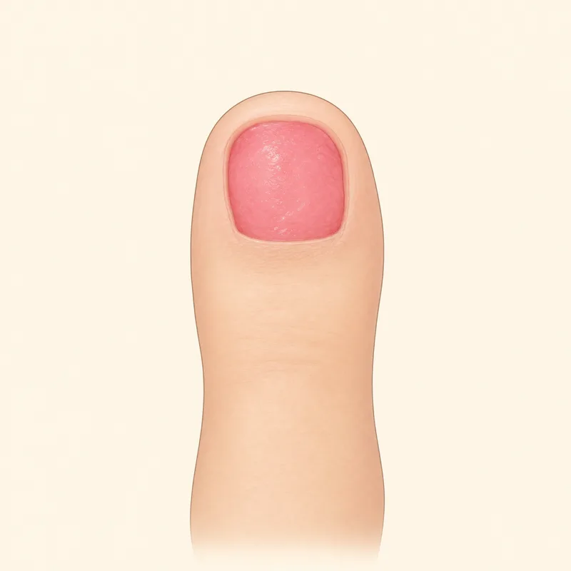
Deep pink to dull red surface
NormalTypically seen: Days 1 to 7
The exposed nail bed itself. Smooth, slightly ridged, moist looking, and sitting a little lower than the skin around it. This is the default appearance and it looks worse under a bathroom light than it is.
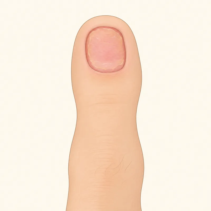
Thin yellow or creamy film
NormalTypically seen: Days 3 to 21
Fibrin, the body's temporary wound cover. Flat, bonded to the surface, and it does not wipe off. This is the number one thing mistaken for pus. Leave it alone.
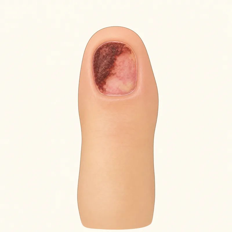
Dark red, brown or black crust
NormalTypically seen: Days 1 to 14
Dried blood, either crusted on top or bruising in the bed from the original injury. It lifts or fades over one to three weeks. New darkening that appears later is the version worth a call.
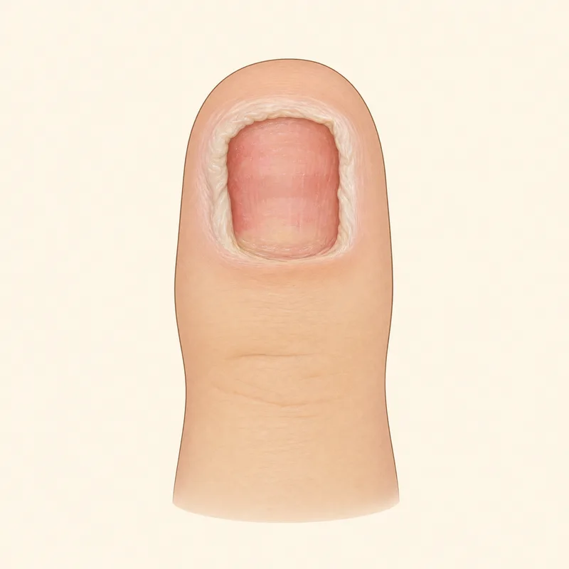
Soft white, wrinkled skin
Keep an eye on itTypically seen: After soaking or a damp dressing
Maceration, meaning waterlogged skin. Harmless briefly, but it means the toe is staying too wet. Shorten soaks to 10 to 15 minutes and dry more thoroughly before re-dressing.
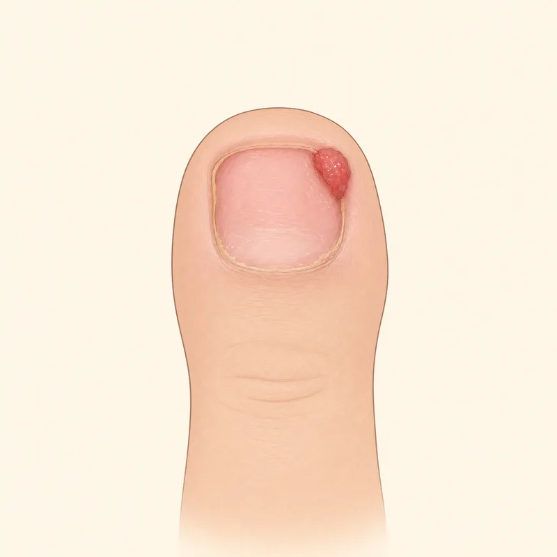
Shiny red mound at the nail fold
Keep an eye on itTypically seen: Weeks 2 to 6
Granulation tissue, sometimes called proud flesh. Glossy, bleeds from light contact, not infected. Most flatten on their own. One that keeps growing or bleeding is a quick office visit.
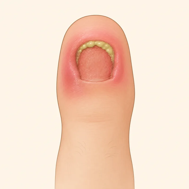
Thick yellow-green drainage
Call your podiatristTypically seen: Any time
Not fibrin. This oozes, reaccumulates after cleaning, often smells, and comes with increasing pain. This is the appearance that actually means infection.
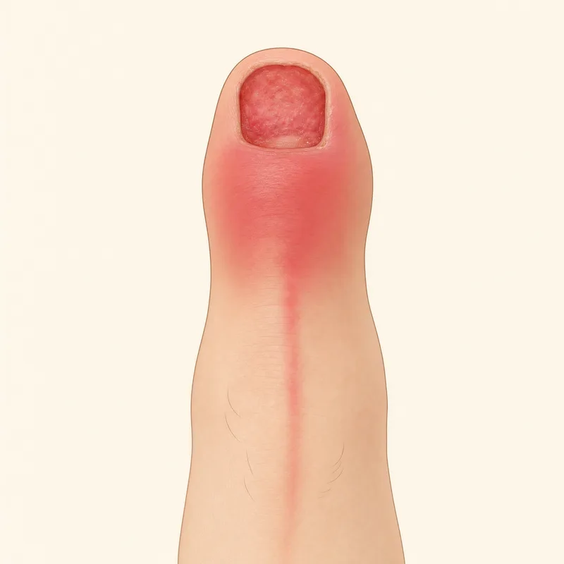
Redness spreading past the nail fold
Call your podiatristTypically seen: Usually days 3 to 7
Normal redness stays as a narrow rim at the fold. Redness that creeps onto the rest of the toe, or streaks running up the foot, needs to be seen, and streaks mean same-day.
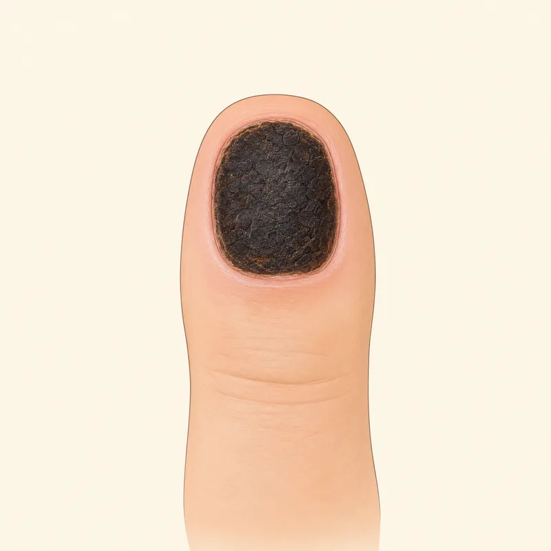
Firm, leathery black tissue
Call your podiatristTypically seen: Any time
Different from a blood crust: dry, hard, and often enlarging. Call promptly, and immediately if you have diabetes, neuropathy, or poor circulation.
Fibrin vs. Pus: The One Distinction Worth Learning
If you take one thing from this page, take this. More patients call about the yellow film than about anything else, and the film is almost never the problem.
Fibrin (normal)
- •A flat film bonded to the wound surface
- •Pale yellow to creamy, sometimes slightly grey
- •Does not wipe away, and should not be scrubbed off
- •No smell
- •Pain is going down, not up
Pus (call)
- •Thick, sits in a pocket or oozes out of the fold
- •Opaque yellow-green, sometimes brownish
- •Wipes away, then comes back within hours
- •Often has a distinct smell
- •Pain, swelling, and redness are increasing
One more clue that separates them without any judgment about color: fibrin appears and then the toe keeps improving. Infection appears and everything else gets worse with it. For the full sign-by-sign comparison, see infection vs. normal healing after toenail removal.
How the Appearance Changes Over Time
Early on the bed is at its most raw looking: wet, deep pink or red, with a crust or two at the edges. Over the next couple of weeks it dulls and dries, the yellow fibrin film comes and goes, and the surface starts to feel less tender to the lightest touch. That is the resurfacing stage, and the change is gradual enough that people who look every day often think nothing is happening.
By the second month the exposed bed usually looks like tough, matte, pale pinkish tan skin rather than a wound. It is often slightly ridged, which is the imprint of the nail bed's natural grooves, and it can be a shade darker or lighter than the surrounding skin for a long time. None of that is a problem.
What happens after that depends entirely on whether the nail root was treated. Our partial vs. total nail avulsion guide explains which version leaves a permanently bare bed and which one grows a nail back, and the regrowth timeline covers what the returning nail looks like month by month.
When Looking Is Not Enough
Appearance is the least reliable of the healing signals, and it is the one people lean on hardest.
A toe can look unremarkable and still be infected, particularly early. It can also look genuinely gruesome and be healing perfectly. The signal that outperforms appearance is direction: pain, swelling, and redness should all be a little better each day from day 2 onward. If any of those three is climbing while you are reassuring yourself that the toe looks okay, trust the direction.
This matters most if you have diabetes, peripheral neuropathy, or poor circulation. Reduced sensation removes the pain warning entirely, so a toe can be significantly infected while looking mild and feeling fine. In that situation, treat any change at all as a reason to be seen. Learn about our diabetic wound care →
Want the week-by-week healing sequence? What happens, and when. Toenail Removal Recovery Timeline →
Was yours an ingrown nail procedure? The border heals differently. Ingrown Toenail Removal Recovery →
Draining for weeks? That is expected after a phenol procedure. Phenol Matrixectomy Recovery →
Frequently Asked Questions
What does a toe look like without a toenail?
Less dramatic than most people brace for. The nail bed underneath is smooth, slightly ridged skin, usually deep pink to dull red in the first days, and it sits a little lower than the surrounding skin so the toe looks flatter at the tip. It is not an open hole and there is no exposed bone or muscle. Within a few weeks that surface toughens and dulls to a pale pinkish tan that looks more like ordinary skin, and from a normal distance the toe mostly looks like a toe with a short or missing nail.
Why is there a yellow film on my nail bed?
That thin yellow or creamy layer stuck to the surface is almost always fibrin, a protein the body lays down over any healing wound as a temporary cover. It is the single most common thing patients mistake for pus. The distinction is that fibrin is a flat film bonded to the wound and does not wipe away, while pus is thick, sits in a pocket or oozes out, and usually comes with increasing pain, spreading redness, and a smell. Do not scrub fibrin off, it is doing a job.
What does an infected toe look like after nail removal?
Infection changes the toe rather than the wound surface. Look for redness spreading beyond the nail fold onto the rest of the toe or up the foot, swelling that is getting worse instead of better after day 2, thick yellow-green drainage that reaccumulates after you clean it, a foul smell, and skin that feels hot. Red streaks running up the foot or ankle, fever, or chills mean you should be seen the same day. Appearance alone is unreliable, so weigh it alongside pain that is increasing rather than decreasing.
Is it normal for the nail bed to look dark or black?
A dark red, brown, or black area in the first week is nearly always dried blood, either a crust on the surface or old bruising in the nail bed from the original injury or procedure. It lifts or fades over one to three weeks. What is worth a call is new darkening that appears later, black tissue with a firm or leathery texture, or a dark area that keeps enlarging, especially if you have diabetes or poor circulation.
What is the shiny red lump at the edge of my nail?
That is granulation tissue, sometimes called proud flesh. It is a mound of new blood vessels and connective tissue that the body overproduces at the nail fold while healing. It looks alarming because it is bright red, glossy, and bleeds from the lightest contact, but it is not an infection and it is not cancer. Most settle flat on their own over a few weeks. If one keeps growing, keeps bleeding, or blocks the nail fold from closing, your podiatrist can treat it in a short office visit.
Will my toe look normal again?
In most cases yes, though the timeline is long. If no matrixectomy was done, a full toenail regrows over about 9 to 12 months and a partial border over 3 to 6, and the new nail often arrives thick, ridged, or slightly discolored for the first months before it evens out. If a matrixectomy was done, a permanently narrower nail or no nail is the intended outcome, and the exposed bed toughens into skin that looks unremarkable once it has matured.
Specialized Wound Care at ASG Foot & Ankle
Our board-certified podiatrists treat slow-healing wounds, diabetic foot ulcers, and post-surgical wounds at our three clinics in Homewood, South Chicago Heights, and Mokena.
See a Podiatrist Near You
ASG Foot & Ankle has offices in Homewood, South Chicago Heights and Mokena. Our board-certified podiatrists accept most insurance. Find the office closest to you:
Want Someone to Actually Look at It?
Comparing your toe to a description online only gets you so far. Our board-certified podiatrists can check it in person at any of our South Chicago suburbs locations, usually the same week. Call us or book online today.